
“Structure and emergence of specific olfactory glomeruli in the mouse”
Potter, S. M., C. Zheng, D. S. Koos, P. Feinstein, S. E. Fraser and P. Mombaerts (2001). Journal of Neuroscience 21:9713-9723.
ABSTRACT
Olfactory sensory neurons (OSNs) expressing a given odorant receptor (OR) gene project their axons to a few specific glomeruli that reside at recognizable locations in the olfactory bulb. Connecting ~1000 populations of OSNs to the ~1800 glomeruli of the mouse bulb poses a formidable wiring problem. Additional progress in understanding the mechanisms of neuronal connectivity is dependent on knowing how these axonal pathways are organized and how they form during development. Here we have applied a genetic approach to this problem. We have constructed by gene targeting novel strains of mice in which either all OSNs or those that express a specific OR gene, M72 or M71, also produce green fluorescent protein (GFP) or a fusion of tau with GFP. We visualized OSNs and their axons in whole mounts with two-photon laser scanning microscopy. The main conclusion we draw from the three-dimensional reconstructions is the high degree of morphological variability of mature glomeruli receiving axonal input from OR-expressing OSNs and of the pathways taken by the axons to those glomeruli. We also observe that axons of OR-expressing OSNs do not innervate nearby glomeruli in mature mice. Postnatally, a tangle of axons from M72-expressing OSNs occupies a large surface area of the bulb and coalesces abruptly into a proto-glomerulus at a reproducible stage of development. These results differ in several aspects from those reported for the development of glomeruli receiving input from OSNs expressing the P2 OR, suggesting the need for a more systematic examination of OR-specific glomeruli.
Keywords:
olfaction; olfactory system; olfactory bulb; glomerulus; sensory neuron; olfactory receptor; odorant receptor; tau; green fluorescent protein; two-photon microscopy; axon guidance
On-line supplemental material
Animation 1: Section series through the olfactory epithelium of a PD2 OMP-GFP mouse.
Same specimen as shown in Fig. 2E-H. The optical sections are taken from superficial to deep. Dendritic knobs are followed by dendrites, cell bodies, axons, and axon fascicles. Only with TPLSM it is possible to see dim features as single axons underneath a layer of extremely bright structures such as the cell bodies.
Animation 2: Section series through the olfactory bulb of an adult OMP-GFP mouse.
Same specimen as shown in Fig. 3 C,D. The optical sections are taken from superficial to deep. The outer nerve layer is followed by the glomerular layer.
Animation 3: Section series through the olfactory bulb of a PD4 OMP-GFP mouse.
The optical sections are taken from superficial to deep. The outer nerve layer is followed by the glomerular layer. The glomeruli are smaller and less well formed as in Animation 2.
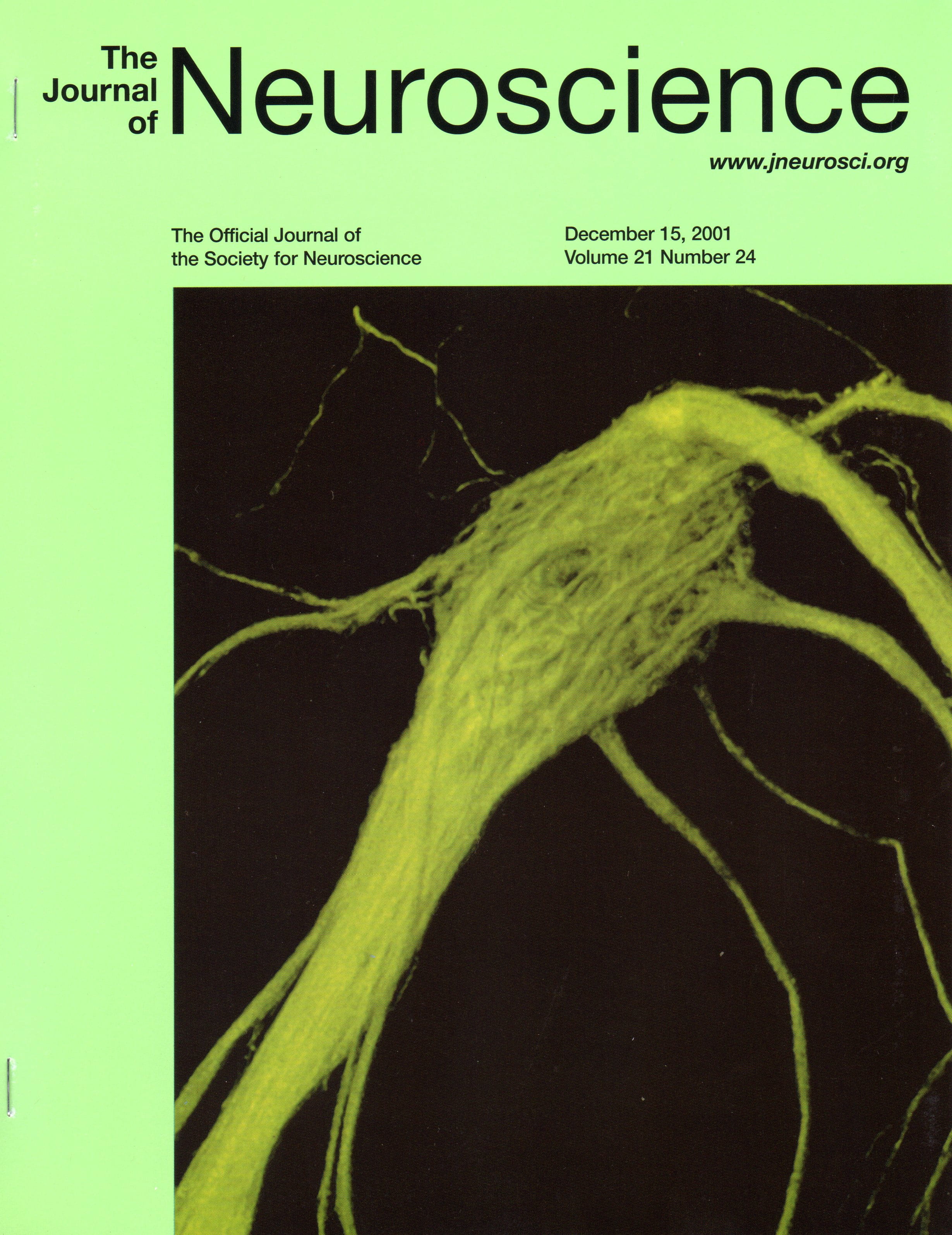
Animation 4: Section series through an M72 glomerulus of a mature M72-IRES-tauGFP mouse.
Same glomerulus as shown in Fig 4C, and on the cover of the Journal. The images are taken from deep to superficial.
Animation 5: Rotating reconstruction of an M72 glomerulus of a mature M72-IRES-tauGFP mouse.
Same glomerulus as shown in Fig 4C. and the cover of the Journal. The rotation provides a better appreciation of the various levels at which axons and axon fascicles approach the glomerulus.
Animation 6: Rocking reconstruction of an M72 glomerulus of a mature M72-IRES-tauGFP mouse.
Same glomerulus as shown in Fig 4D. It is the lateral glomerulus in the left bulb of the same mouse as the glomerulus shown in Animation 5.
Animation 7: Rocking reconstruction of an M72 glomerulus of a mature M72-IRES-tauGFP mouse.
Same glomerulus as shown in Fig 4E. Numerous smaller fascicles arrive from directions spread over 360 degrees.
Animation 8: Rocking reconstruction of an M72 glomerulus of a mature M72-IRES-tauGFP mouse.
Same glomerulus as shown in Fig 5D. Most fascicles arrive from down under, but one fascicle (top right) approaches the glomerulus from a very different orientation.